
Animal Cell Diagram
In this article, you will be learning about the topics mentioned below:
What is cell?
Types of cell
Animal cell definition
Animal cell diagram
Parts of an animal cell with functions
Different types of animal cell
Plant cell vs animal cell
Animal cell vocabulary
Animal cell FAQs
Animal cell Quiz
What is cell?
Types of cell
Eukaryotic cells:
- Eu + Karyotes = eu means true, and Kary means nucleus.
- Have a ‘true’ nucleus surrounded by a nuclear membrane called the nuclear envelope where DNA is stored.
- Larger and more complex than prokaryotic cells.
- Found in plants, animals, fungi, and protists.
- Can be unicellular and multicellular.
Prokaryotic cells:
- Pro + Karyotes = pro means before, and Kary means nucleus.
- Smaller and simpler than eukaryotic cells.
- Single-celled organisms (bacteria and archaea) are made up of a single prokaryotic cell.
- Lacks a nucleus and membrane-bound organelles.
Animal cell definition
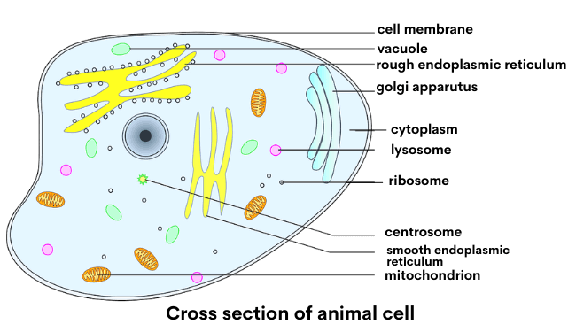
Animal cell diagram

If you compare the animal cell diagram with a plant cell diagram, you can easily distinguish them by observing the presence or absence of a cell wall.
Note: This animal cell diagram provides information on the internal structure of most animal cells. It does not represent any particular type of cell.
Parts of an animal cell
In this section, we will be discussing the several parts of an animal cell with their functions.
The parts of an animal cell are called organelles, that do a variety of things, including:
- Process and release energy
- Destroy and digest materials
- Replicate genetic information
The organelles found in most animal cells include the nucleus, cell membrane, cytoplasm, mitochondria, ribosomes, lysosomes, vacuoles, centrosome, endoplasmic reticulum, and Golgi apparatus.
Cell Membrane
This is the outer barrier of the animal cell. It controls what goes in and out of a cell. The cell membrane, also known as the plasma membrane, is semipermeable, which means it can restrict some stuff to cross it.Cytoplasm
Cytoplasm is a jelly-like substance inside the cell membrane, containing all the parts of a cell such as mitochondria, ribosomes, endoplasmic reticulum, Golgi apparatus, lysosomes intermediate filaments, microfilaments, microtubules, vesicles.
It is made up of mostly water, but many other substances are dissolved in it. The cytoplasm is responsible for maintaining the cell’s shape.
Mitochondria
Mitochondria, a rod-shaped or oval-shaped organelle, is the ‘POWERHOUSE’ of the cell. The primary responsibility of Mitochondria is to generate energy for the cell. They produce energy in the form of Adenosine triphosphate (ATP).Nucleus
The nucleus is the “brain” or “control center” of a cell, a spherical-shaped organelle located majorly at the center of an animal cell.
It contains deoxyribonucleic acid (DNA), which is the code for genetic information.
The nucleus also includes the nuclear envelope, nuclear lamina, nucleolus, chromosomes, and nucleoplasm.
The primary function of the nucleus is to control the cell’s growth and reproduction.
Ribosomes
Ribosomes are the “factory of protein making” in the cell. They are found floating around in the cytoplasm or bound to the endoplasmic reticulum. Ribosomes receive instructions from the hereditary material to make protein for the cell.Lysosomes
It is the digestive system of the cell. Mainly, it is responsible for digestion, excretion, and cell renewal process. They have chemicals to break down food, cell waste, and the foreign particles that enter the cell, such as bacteria and viruses.Vacuoles
Vacuoles act like “the storage unit of the cell” because they store water, food, and waste.Centrosome
The centrosome serves as the main microtubule-organizing center (MTOC) of the animal cell and provides a structure to the cell.Endoplasmic reticulum
Endoplasmic reticulum acts like “the transport facility for the cell” because it helps produce and transport proteins and lipids.Golgi apparatus
Golgi apparatus, also known as the Golgi body, act like “the packaging, sorting, and distributing center of the cell.” They sort nutrients (proteins and lipid molecules) processed by the endoplasmic reticulum, then package and distribute them out of the cell.Different types of animal cell
Skin Cells
Skin cells include melanocytes, keratinocytes, Merkel cells, and Langerhans cells.
Blood Cells
Blood cells are responsible for carrying oxygen throughout the body as well as collecting carbon dioxide. Apart from carrying oxygen, these cells also carry hormones, enzymes, and vitamins to different parts of the body.
Blood cells include RBC (red blood cells) and WBC (white blood cells that help fight infections).
Nerve Cells
Nerve cells, also called neurons, are found in the nervous system. Did you know? The human brain has 100 billion neurons and 10- to 50-fold more glial cells (source).
“The African elephant brain, weighing 4618.6 g (without the olfactory bulb), about 3 times heavier than the human brain, holds a total of 257.0 billion neurons, also 3 times more than the average of 86 billion neurons found in the human brain. source”
The nerve cell sends and receives electrical signals. The neuron is made up of a cell body called dendrites and a threadlike structure named axon. Dendrites bring electrical signals to the cell body, and axons take information away from the cell body. (For detail)
Muscle Cells
Muscle Cells, also known as Myocytes, Muscle fibers, are responsible for moving an organism’s limbs and organs. It includes skeletal, cardiac, and smooth muscle cells.
Fat Cells
These cells are also known as adipocytes or lipocytes and are responsible for storing fats and other lipids. White fat cells and brown fat cells are the two common types of fat cells in animals.
Plant cell vs. Animal cell diagram

Differences between Plant cell and Animal cell
Plant cell
- typically rectangular or cubic
- The cell wall is present
- have chloroplasts
- Centrosomes are absent
- store energy in the form of starch
- have plastids
- usually have one or more large vacuole(s)
Animal cell
- typically round or irregular shaped
- The cell wall is absent
- do not have chloroplasts
- Centrosomes are present
- store energy as complex carbohydrate glycogen
- do not have plastids
- may have smaller vacuoles
Animal cell vocabulary
Cell membrane – outer barrier of the animal cell, controls what goes in and out of a cell
Nuclear envelope – surround nucleus
Nucleus – The “brain” or “control center” of a cell, contains the code for genetic information.
Nucleolus inside of nucleus
Nuclear sap fluid in nucleus
Cytoplasm – The jelly like substance inside of the cell, not including the nucleus.
Mitochondria – ‘POWERHOUSE’ of the cell, generate energy for the cell.
Golgi complex – The packaging, sorting, and distributing center of the cell.
Ribosomes – “Factory of protein making”, make proteins for the cell
Lysosomes – “the digestive system of the cell”, help in digestion, excretion & cell renewal process.
Endoplasmic reticulum – “The transport facility for the cell”, helps produce & transport proteins and lipids
Microbody cotain enzymes look like small vacuoles
Vacuoles – “the storage unit of the cell”, store water, food, and waste
Centrioles – function during cell division.
Animal cell diagram FAQs
What is an animal cell?
An animal cell is a eukaryotic cell that lacks a cell wall.
Do animal cells have a cell wall?
No, animal cells do not have a cell wall.
What are the types of animal cells?
The different types of animal cells are skin, nerve, muscle, blood, skin, and fat.
What structures are present in an animal cell but not in a plant cell?
- Cell walls are present in plant cells but absent in animal cells.
- Chloroplasts are present in plant cells absent in animal cells.
- Centrosomes are absent in plant cells but present in animal cells.
- Plant cells have plastids, but animal cells do not contain them.
What does the vacuole do in an animal cell?
Vacuoles act like “the storage unit of the cell” because they store water, food, and waste.
What does the rough endoplasmic reticulum do in an animal cell?
To produce proteins is the main function of the Rough Endoplasmic reticulum.
Test your Knowledge on Animal Cell Diagram
It is time to put your knowledge to the test by answering a few questions. Click ‘Start Quiz’ to begin!